
The human brain and the human eye cooperatively translate light into color. Light receptors are present in the eye, which transmits the messages to the brain. These light receptors are present within the human eye to produce the confidential sensation of colors. Newton perceives that color is not inherent in objects. Instead, the surface of objects soaks up some of the colors and reflects unabsorbed colors. This means that the surfaces reflect specific wavelengths and absorb all the others. When an object reflects all the wavelengths, it appears as white, and on the other hand, it appears black when it absorbs the entire wavelength.
Color Spectrum, Cones, and Rods
If we talk about the colors spectrum, some colors are additive primary colors of the color spectrum: red, green, and blue. By varying the amount of these primary colors of the color spectrum, which are Red, Green, Blue, all of the other colors in the visible spectrum can be produced. While by mixing a well-balanced amount of these additive primary colors of the color spectrum, we can also create pure white.
Our eye contains two types of photoreceptors, which are rods and cones. The rods allow us to see at in low light levels, whereas the cones allow us to see in high light levels. In each eye, there are 120 million of each of them. Because of the rods and cones, our peripheral vision is less sharp and colorful as compared to our front on vision. The rods are mostly strenuous around the edge of the retina. Most of the time, rods transmit black and white information to the brain, and since rods are much sensitive to dim light than cones, we lose most of the colors vision in darkish light, and our peripheral vision is can be less colorful. When we enter a dark room, the rods help our eyes to adjust to the low light intensity level inside that dark room. The light enters into the eye and then to the retina placed on the backside of the human eye, and we distinguish violet from blue and know that the rose is red only because of the specialized cells in our eyes which are called cones. Cones are responsible for color in our vision.
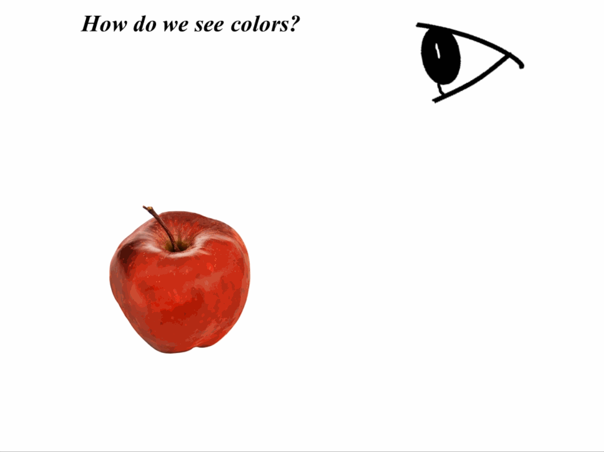
When the light hits an object, here, as an example, we say the object is an apple (as shown in Fig. 1). This object soaks up some part of the light and reflects the rest of the light. Which part of these wavelengths are absorbed and reflected depends on the properties of the object. If we talk about the ripe apple, then wavelengths of 620-633 nanometers bounce back, and these wavelengths are of red light. When we look at an apple, some part of the light is reflected. The wavelength of this reflected part determines the specific color we see. The light wave reflects off the object’s peel and moved to the light-sensitive retina located at the backside of our eye where cones have come in and these cones are one type of photoreceptor, and these tiny cells in the retina of our eye respond to the light. This is called the fovea centralis, which is about 0.3-millimeter spot on the retina. Most of us have around 5-7 million cones, and almost all of them are concentrated in this 0.3-millimeter spot.
Not all of these millions of cones have the same behavior. We talked before about the additive primary colors red, blue, and white, around 64 % of these cones highly respond towards the red light while around 34 % respond most by green light and the rest of the 2 % strongly respond to the blue light. Scientists’ estimation is that some humans can differentiate the millions of colors but this is not true for every human. Some people can see 100 million colors, but on the other hand, some people can see only a few hundred colors.
Types of color blindness and their causes
The deficiency in color vision is called color blindness. Color blindness happens when one type of cone does not work are we can say completely missing from the retina. As we learned, there are different types of cones that mean that there are also different types of color blindness. Color blindness depends on which type of cone is missing or not working in our eyes.
If red cones or completely missing or not working is called protanopia, if green cones are missing or not working, this is called deuteranopia. Both conditions of color blindness are usually referred to red-green color blindness. This red-green color blindness makes it very difficult to distinguish between different colors such as shades of red, orange green, and yellow and this type is the most common type of color blindness
People who have protanopia are less sensitive to red light. And especially rainbow colors we know about that a person who has protanopia might possibly see it as a blue and yellow color. And the other hand people who have deuteranopia are less sensitive to green light. The person with this type also sees the color yellow and blue but the color will be different. The person with this problem might see the rainbow-like this.
One another type of color blindness is tritanopia. The people who have this problem cannot differentiate between the yellow and blue colors. Because of these colors, this is also called yellow-blue color blindness. The issue of tritanopia happens when the blue color cone missing or does not work. Although people with this problem are rare and these people have difficulty differentiating between the colors such as blue from green and yellow and purple. These people might possibly that see the rainbow as shades of pink green and red. They see the rainbow in this form
Another form of human eye color blindness is Achromatopsia, although it’s also very rare it happens. Achromatopsia have further two types one is incomplete Achromatopsia and another one is incomplete Achromatopsia. In Incomplete Achromatopsia, two types of cones are missing out of three types of cones and the brain needs at least two types of cones to compare signals and identify the different colors. So that is the reason most people with this problem have limited color vision. While incomplete Achromatopsia all type of cones is missing or does not work people with this problem see the colors but entirely in shades of grey. People with complete Achromatopsia may see the rainbow-like this the rainbow-like this
If we talk about the causes of color blindness in the human eye it is observed that most of the color blindness problems are happening are the result of genetic mutations. Some of the genetic mutations cause cones cells to only partially work, these all reasons milder to form color blindness of someone. Other genetic mutations cause completely missing cone cells. Other than genetic mutations color blindness can also be the result of other different reasons such as chronic illness or taking certain different medications or brain damage.
On another side of the spectrum, with the different researches and with the different theories Researchers also observe and discovered that it may be possible that around 12 percent of women have four types of cones in their retinas it’s this is called tetrachromacy and that human may have this is called tetrachromat. Scientists observe and suggested that these types of people may see up to hundreds of millions of different colors which the average person cannot see. Human Eye is a very special organ of the human body it is a specialized sense organ that is capable of receiving visual images which are carried to the brain from the eye.
Anatomy of the Human eye
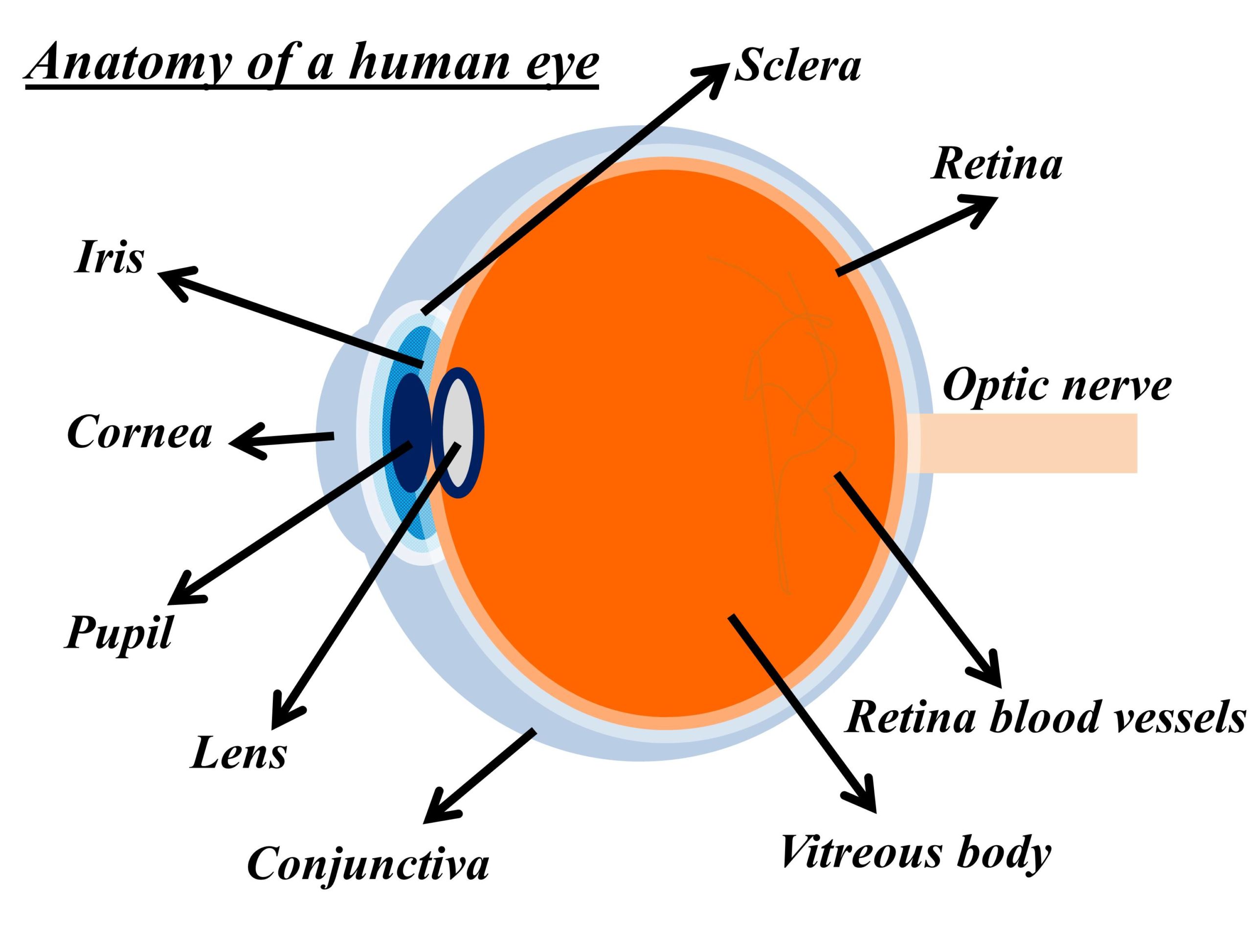
The human eye and its structure are shown in Fig. 2.
Structures auxiliary to the
The orbit
Our eye is enclosed into the orbit or socket made by the portion of several bones of the skull. This orbit forms a four-sided pyramid. Because of this, our eye is protected from mechanical injury. The flour of this orbit is made up a part of the maxilla.
The eyelid
It is essential that the next surface of the bandage, the cornea is moist. It is obtained by the eyelid, which regularly stir up secretions from the lacrimal tract and other glands upon awaking and cover the eyes during sleep and prevent evaporation. In the blink of an eye, the eyelid has the added function of preventing foreign body injuries.
The conjunctiva
The conjunctiva covers the eyelids and then attaches to the eyelid’s surface, forming an outer cornea towards the front of the eye and ending in the transparent area of the eye, the cornea.
The fibrous layer
The fibrous layer, which gives mechanical stability to the eyelid, is composed of thick, relatively rigid tarsal plates, which directly limit the eyelid opening, and the much thinner eyelid fascia or connective tissue sheet; the two together are called the orbital septum.
The muscles of the lids
Eyelid closure is achieved by contracting the orbicularis muscle, a single oval sheet of muscle that extends from the forehead and face regions and surrounds the orbit to the eyelids. It is divided into orbital and eyelid portions, and it is essentially the portion of the eyelid within the eyelid, that causes the eyelid to close. The orbicularis and odor are striated muscles under voluntary control.
The front part of the eye contains the following.
Iris
It is the colored part of the eye. The color may vary from human to human.
Cornea
It is the clear dome-shaped covering over the iris of the eye.
Pupil
It is the black circular opening inside of the iris to let the light inside of the iris.
Sclera
The portion of the eye covered in white color is the Sclera.
Conjunctiva
It is the thin tissue layer covering pur entire eye with an exception of cornea.
Further readings
If you liked this post, you might be interested in reading the following posts.
- Construction, working, and new technologies of OLED TV
- Laser and its applications in medicine and technology
- LED light, its construction, types and colors, power, life, and technology
- Optical fiber design and applications

- Radiation therapy for cancer treatment and its side effects

- Refractive Index of a material and material properties
- What are Metamaterials and Metasurfaces?
- Thin film interference of light
- Solar cells, their construction, and working
- Solution of complex multilayer thin films by Matrix Method
- From solar cells to solar panels to photovoltaic (PV) system
- Shading on solar panels and its types
- The function of Bypass diodes in increasing power output
- Climate change leading to global warming
- Components of a solar PV system
I am not rattling fantastic with English but I get hold this rattling easygoing to interpret.